Lasik or lenses | See without glasses
Basics of refractive surgery
|
Ametropia and its correction The surface-curvature of the cornea, the refractive power of the lens and the length of the eyeball just and their proportion. If the dimensions of these three elements are perfectly adjusted, the light will be focused directly on the retina and one will see sharply (=emmetropia). In many cases, however, there are misproportions in the relation of these dimensions. We are speaking of ametropia or a "refraction anomaly". |
|
Myopia (shortsightedness) |
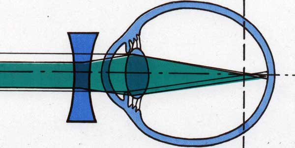 The optical correction happens by reducing the refractive power and consequently shifting the focus further back. The necessary factors are specified on glasses or contacts (e.g. -4,5 dpt). The optical correction happens by reducing the refractive power and consequently shifting the focus further back. The necessary factors are specified on glasses or contacts (e.g. -4,5 dpt). |
|
|
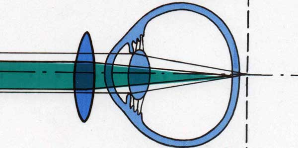 However, this ability will fade age, which everyone will eventually experience when looking objects that are close. The optical correction will occur by raising the refractive power with a converging lens, thus shifting the focus further to the front. The necessary factors are specified on glasses or contacts (e.g. +4,5 dpt). However, this ability will fade age, which everyone will eventually experience when looking objects that are close. The optical correction will occur by raising the refractive power with a converging lens, thus shifting the focus further to the front. The necessary factors are specified on glasses or contacts (e.g. +4,5 dpt). |
|
Astigmatism is commonly caused by the distortion of the cornea but it can also have its origin in the lens. This has to be checked in the preliminary investigation and is important for the choice of surgery. |
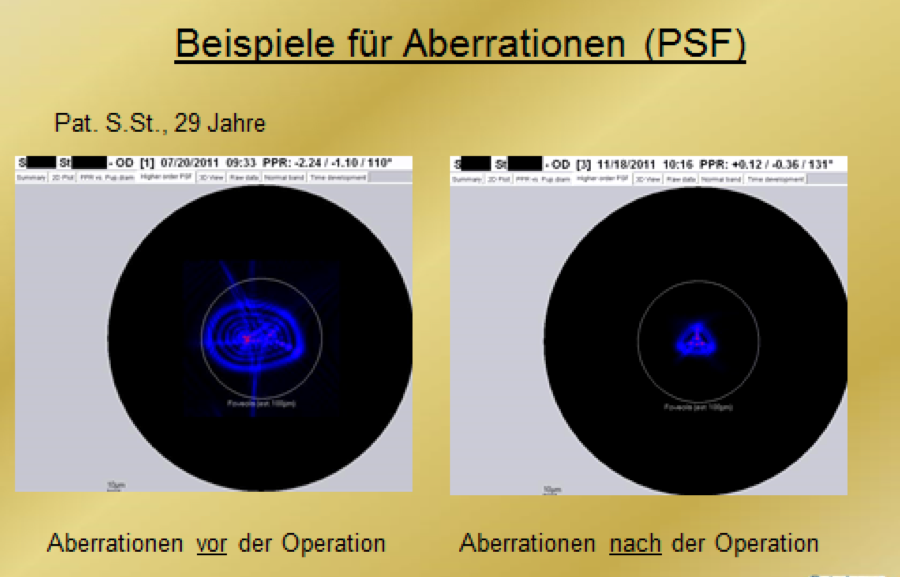
| Aberrations - what are they? Only in the case of an ideal optical system, the optical pathways apply for emmetropic as well as ametropic eyes, but the human eye exhibits aberrations: disortion of images, caused by irregularities in the optical system. We differentiate between chromatic and spherical aberrations. Although these were discovered decades ago, we were not able to measure them in the eye. Due to the development of machines to measure aberrations, new possibilities are being established concerning diagnostics as well as treatment of several kinds of sight disorders (chromatic aberrations remain unconsidered). » see also below (at Diagnostics) |
Diagnostics
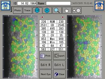
As always, we first have to determine whether surgery is reasonable or not. Moreover, a refractive operation is mostly done on a healthy eye. Thus the medical history, individual psychological stress and the motivation of the patient play big roles. In addition to the normal ophthalmological functional and organic inspection, special diagnostics are necessary for refractive surgery. These are:
|
|
|
|
|

That's why - in every preliminary LASIK investigation - we take data from all of the three devices that are available to us so that we get to most accurate data possible for us to gather.
|
- Pupillometry
…for the determination of the diameter of the pupil influenced by dusk or dawn. It is important for the evaluation of the potential of experiencing glare in a dark environment (e.g. with Plusoptix) - Ultrasound-biometrics
...for the measurement of the entire eye via ultrasound, making it possible to evaluate the dimension of the individual parts of the eye and for the calculation of implants - B-scope-sonography
...for recognizing and judging structures in the eye - IOL-Master (Zeiss)
...for noncontact photo-optical measurement of the eye, especially for the calculation of the lens-implants - Analysis of tear film(see also Tear labratory)
Methods of treatment

- Overview
- LASIK
- Femto-LASIK
- ReLEx smile
- PRK & PTK
- Lens implants
- Clear lens extraction
- Astigmatismus-correction
- Varia
Overview
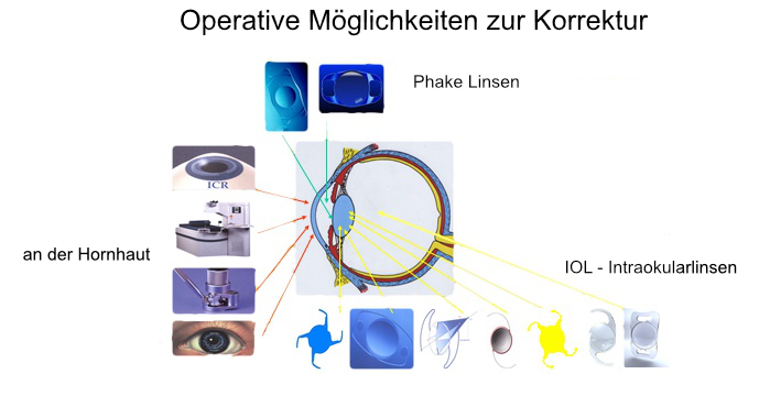
Refractive corrections on the cornea can always be done by replacing the lens or with an extra lens in between the cornea and the natural lens.
LASIK
LASIK (laser-in-situation-keratomileusis) is probably the most well known and most used method of refractive surgery. In combination of microsurgery-like cutting technique and removal of tissue by the Excimer-Laser, quick and exact results can be reached.
Special andvantages of LASIK:
- wide area of correction
- fast regeneration of visual acuity
- pain-free healing phase
- stable results
What is going to happen?
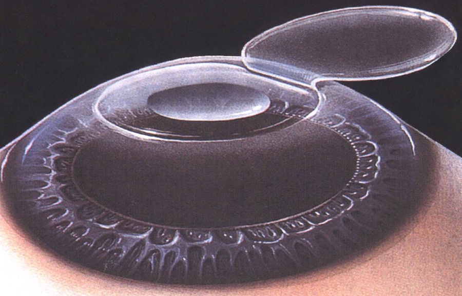
After covering the lids with anesthetic eye drops, first there will be a dissection of the cornea-lamella by microtome or femtosecond laser, which will be lifted, so that the corneal tissue below can be modeled suitably. When the lamella is back in place, it will suck itself onto the eye and will strongly reconnect to it in a matter of days. The pure laser treatment usually takes less than a minute and the whole operation takes less than 15 minutes. The laser procedure is completely painless and invisible for the patient.
After one to two days very good visual acuity will develop and become stable within the next month. For a few days after the operation antibiotic and moisturizing eye drops should be used and, if necessary, a protective shield should be worn while sleeping.
In the first few days, most patients will feel only a slight sensation of a foreign body in the eye. Higher glare and lower visual acuity during the night are often observed in the first weeks and especially can occur for people with relatively large pupils and higher corrections. Even though, for a majority of patients, the exact correction that has been requested is reached after the treatment, another correction can become necessary. Usually after some months.
LASIK is a very safe and scientifically approved method. Complications normally do not occur, and when they do they can mostly be corrected with another treatment. We also provide preservative-free antibiotic eye drops for pre-operative pretreatment during the preliminary examination.
BEFORE SURGERY
Three days before the operation, eyelid-makeup should not be used and on the day of the operation one should not use perfume.
The operation is done on an outpatient basis. The patient should be picked up because he/she is not allowed to drive for 24 hours after surgery. In addition, because of the general medication (ibuprofen and dormicum), no machines can be operated and no meaningful decisions should be made.
The operation is done on an outpatient basis. The patient should be picked up because he/she is not allowed to drive for 24 hours after surgery. In addition, because of the general medication (ibuprofen and dormicum), no machines can be operated and no meaningful decisions should be made.
AFTER SURGERY
An eye which has been recently operated on requires careful handling. In the days after the operation the concerned eye should not be rubbed. The patient also should not be too physically active, should not visit a public pool and should not apply any makeup. Eye drops, eye ointment and other medication(s) should be used regularly or taken as ordered. All follow-up examinations are essential. For our patients, we usually recommend: one day after surgery, one week, one month, three months, one year.
A laser surgery of the cornea has effects on subsequent measurements of the eye. Therefore, the patient should tell the ophthalmologist about the any such laser operation prior to measurement of ocular tension or pre-examination for cataract surgery. If any abnormalities occur that may indicate complications, the doctor should be contacted immediately. Our surgeon will always be available for you.
POSSIBLE COMPLICATIONS OF A LASIK-TREATMENT
- If left untreated, infections can lead to corneal clouding and permanent reduction of visual acuity. They are extremely rare in Central Europe.
- Deviation of the cutting quality
- Under certain anatomical conditions of the eye, certain technical problems can lead to difficulties in the preparation of the corneal flap. This may result in interruption or rescheduling of the procedure, or another method may be advisable.
- Healing disorders (or delays) can cause asymmetric surfaces and thus irregular astigmatism which usually nomalize after a few months or, if necessary, need to be corrected.
PERSONALIZED TREATMENTS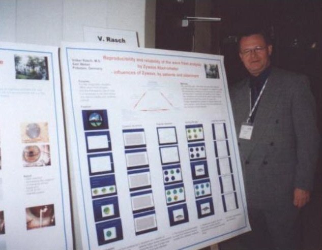 The correction of spherical aberrations using the laser treatment was started years ago. At first, there was euphoria but it needed to be critically reviewed. As a result, we examined various factors influencing aberrations in the eye. Our results from this "world's first large-scale screening of this issue" were published at the ESCRS 2001 Cannes Congress and honored with the Congressional Prize. The correction of spherical aberrations using the laser treatment was started years ago. At first, there was euphoria but it needed to be critically reviewed. As a result, we examined various factors influencing aberrations in the eye. Our results from this "world's first large-scale screening of this issue" were published at the ESCRS 2001 Cannes Congress and honored with the Congressional Prize. |
|
|
|
Femto-LASIK
Excimer laser treatment after flap preparation with femtosecond laser
The difference between the LASIK and the Femto-LASIK or iLASIK is found in the preparation of the coverage (Flap), which must be flipped aside during the treatment to make the desired correction in the cornea with the excimer laser.
The femtosecond laser achieves a separation of the corneal lamellae through extremely short laser pulses (in the femtosecond range) through the smallest "explosions" in a precisely defined corneal depth. The result is a cut with great precision. The advantages of this method are:
- more accurate cut and depth
- less mechanical parts during preparation
- Thinner slats ("flaps") are possible. This is especially useful at higher diopters.
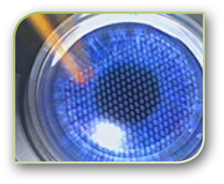
But - femtolaser does not necessarily mean femtolaser.
In October 2008 we decided in favor of the most modern and the safest device: the IntraLase 60 kHz. We have been able to significantly improve the quality of the cut. But even with this device, the development did not come to a standstill. From May 2009, the IntraLase femtolaser with 150 kHz will be available: a module of iLASIK technology. As studies have shown, the cuts are even smoother and more precise. Also, the treatment time is reduced by half - less stress for the patients, less strain on the eye.
WAS IST ILASIK?
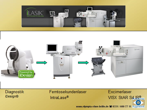
iLASIK ™ describes the combination of the most advanced laser and diagnostic equipment, the IntraLase® FS femtosecond laser, the VISX STAR S4 IR® excimer laser, and the iDesign aberrometer.
Extraordinary technology with every step
The iLASIK ™ system provides a fully integrated, fully customized treatment, using the most advanced technologies in each step..
Step 1 - Creating your personal visual profile
Step 2 - Preparing the iLASIK ™ Flap
Step 3 - Correcting your vision defect with the VISX STAR S4 IR® Excimer Laser
ReLEx smile
For the correction of refractive errors, the individualized femto-LASIK has been regarded as the best scientifically recognized procedure for more than 10 years. The ReLEx smile technology takes a different approach: A narrow opening is prepared by means of a femto laser to prepare a 3D slice (lenticle) inside the cornea (stroma) and remove it via the opening. This lenticule, corresponds in volume and shape to the amount of tissue of your ametropia to be corrected. The stability of the cornea is largely preserved and the production of the tear film is less disturbed. Therefore, ReLEx smile may also be more appropriate for patients with dry eyes or contact lens intolerances.
PRK & PTK
PRK A PRK (photorefractive keratectomy) may be useful if, for example, a LASIK is contraindicated because of too little thickness of the cornea, or in the case of superficial corneal scars. These can be simultaneously reduced during the treatment. With the PRK, e.g. Ametropia with a myopia can be corrected to -5 dioptres as well as a low-grade astigmatism.
In contrast to LASIK, no cut is made in the tissue. Only the corneal epithelium is removed and then the tissue removal is done from the outside. For a few days, the patient wears a so-called therapeutic contact lens.
- Complications
Pain and burning eyes as well as increased tears often persist after the procedure. An eye dryness can also occur. Scars sometimes form, but usually disappear within months. Therefore, vision may be somewhat limited during this time. Especially in the first few months, there might be an increased glare sensitivity. The visual defect can be under- or over-corrected. Also, in very rare cases, the ablation site is not centered exactly so that distorted and blurred images may occur. The procedure can rarely lead to recurrent corneal erosion. The open cornea also rarely causes infections and extremely rare corneal ulcers. In such cases, it could lead to permanent vision deterioration, to blindness or loss of the eye. Also allergic reactions can not be excluded.
Hinweis: Dieser Abschnitt kann nur einen kurzen Abriss über die gängigsten Risiken, Nebenwirkungen und Komplikationen geben und erhebt keinen Anspruch auf Vollständigkeit. Das Gespräch mit dem Arzt kann hierdurch nicht ersetzt werden. - Forecast
In most cases, the PRK can be used to correct the ametropia, provided that the presupposed dioptric values have not previously been exceeded. However, it can not be ruled out that seeing does not get better or even worse. In about 5% of cases, glasses or contact lenses must still be worn. A presbyopia, in which the elasticity of the lens decreases, can not be satisfactorily corrected. Correction attempts have been in trial for years. A disadvantage of PRK is that scarring can form in the cornea.
Most of the time the day after laser surgery visual improvement without correction is noticeable. The end result can only be fully assessed after several months, during which time vision may still vary.
LASEK and Epilasik are operationally equivalent to a PRK. We will gladly inform you about the differences on our information evenings or in a personal consultation.
PTK
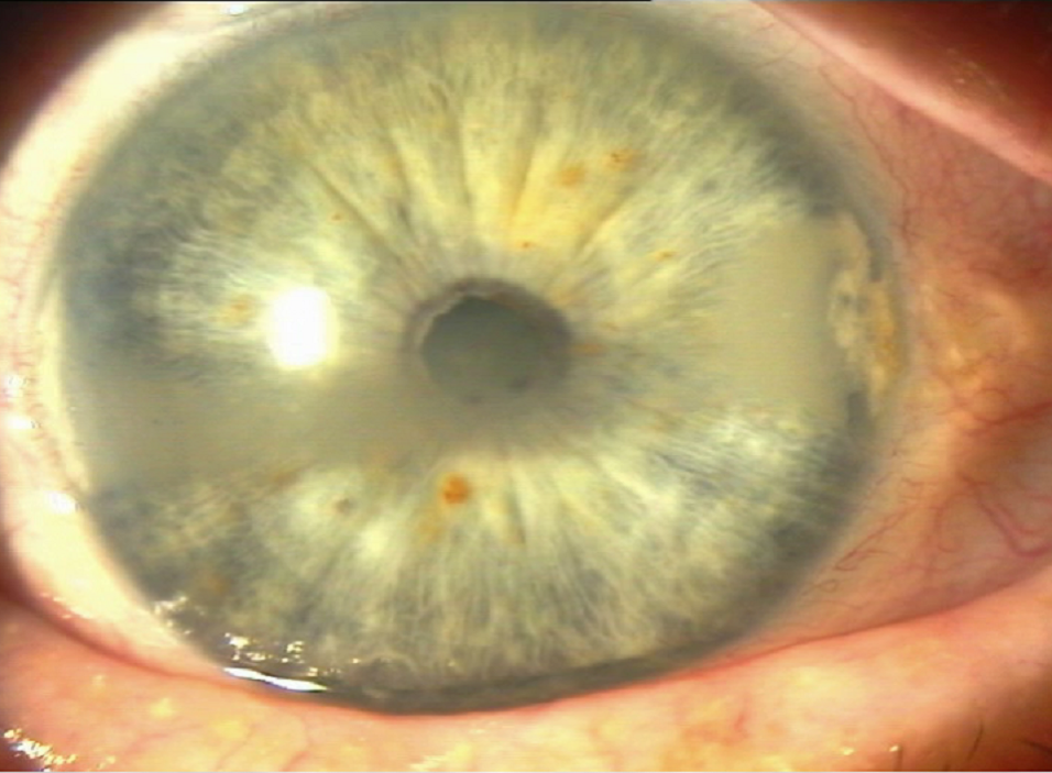
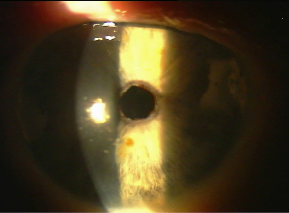
If the removal or reduction of corneal opacities is the goal in the removal of the tissue "from the outside", one speaks of a PTK (Photo-Therapeutic Keratectomy). The indications can be very different. It is also the drug of choice for recurrent corneal erosion. The PTK is in many cases covered by public health insurance. It then will not be done by the eye clinic, but by the practice of Dr. Rasch and his colleagues. (see aslso "PTK-treatment")
Bandförmige Hornhautdegeneration
nach der PTK
Lens implants
Lens implants can either be inserted into the eye (Phake lenses, P IOL) or replaced after removal of the lens (intraocular lenses = ICL).
Phake anterior chamber lens (Verisyse, Artisan)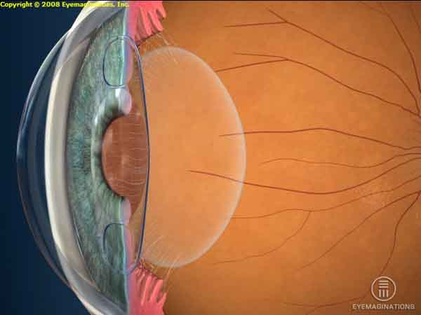 The desired refraction change may also cause a lens implanted between the cornea and the iris into the anterior chamber. It can be attached to the iris or resiliently supported in the chamber angle. With bifocal optics, the presbyopia can be corrected as well. The desired refraction change may also cause a lens implanted between the cornea and the iris into the anterior chamber. It can be attached to the iris or resiliently supported in the chamber angle. With bifocal optics, the presbyopia can be corrected as well. |
Possible complications: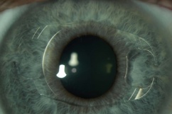 Loosening of the lens and contact with the cornea may cause opacification of the cornea. Loosening of the lens and contact with the cornea may cause opacification of the cornea. |
Intraocular contact lens (VISIAN ICL)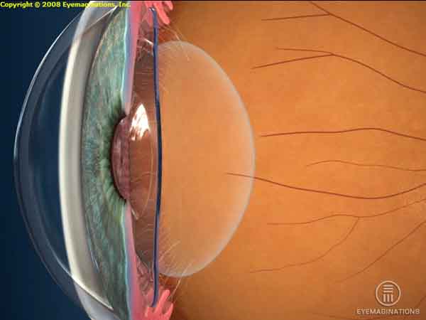 The ICL is implanted in the eye and remains there permanently to change the refractive power of the eye. The lens is made of well-tolerated materials so that virtually no repulsion reactions occur. The ICL is implanted through a 2.5 mm small incision and placed behind the iris and in front of the eye lens. The operation is performed on an outpatient basis under local anesthesia and lasts about 20 minutes. The implantation of an ICL is a reversible method, meaning the contact lens can be removed at any time. The ICL is implanted in the eye and remains there permanently to change the refractive power of the eye. The lens is made of well-tolerated materials so that virtually no repulsion reactions occur. The ICL is implanted through a 2.5 mm small incision and placed behind the iris and in front of the eye lens. The operation is performed on an outpatient basis under local anesthesia and lasts about 20 minutes. The implantation of an ICL is a reversible method, meaning the contact lens can be removed at any time. |
Possible Complications: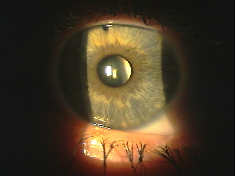 The contact of the ICL with the body's own lens can lead to clouding of the natural lens. The contact of the ICL with the body's own lens can lead to clouding of the natural lens. |
Clear lens extraction
|
(compare "Cataract surgery") |
Astigmatismus-correction
|
In principle, corrections can be made by interventions on the cornea, by lens exchange (for a toric IOL) or by means of a toric phakic IOL. If the cylinder values for a PRK or LASIK are too high, so-called arcuate incisions, which means arcuate sections placed vertically in the cornea, reduce the corneal astigmatism to such an extent that thereafter a PRK can give good results. |
Varia
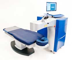 The LaserSoft of the company Katana Technologies is a so-called solid-state laser. It is one of the most advanced and sophisticated systems in refractive surgery. It does not require gas and has other advantages along with the world's smallest 0.2 mm spot size: less corneal temperature and less pain after water treatment. The tissue removal is thus independent of the moisture of the tissue. With a pulse rate up to 4 KHz, the system is considerably faster than all excimer lasers. The very fast video eye tracker is used to track the eye movements and is suitable for the treatment of fine irregularities of the cornea. The "LaserSoft Intelligent Software" allows corrections for myopia, astigmatism and hyperopia and over topolink and ocular wavefront measurement, such corrections as the one of presbyopia are possible. Our clinic has been working with Katana since the development of this new technology. The LaserSoft of the company Katana Technologies is a so-called solid-state laser. It is one of the most advanced and sophisticated systems in refractive surgery. It does not require gas and has other advantages along with the world's smallest 0.2 mm spot size: less corneal temperature and less pain after water treatment. The tissue removal is thus independent of the moisture of the tissue. With a pulse rate up to 4 KHz, the system is considerably faster than all excimer lasers. The very fast video eye tracker is used to track the eye movements and is suitable for the treatment of fine irregularities of the cornea. The "LaserSoft Intelligent Software" allows corrections for myopia, astigmatism and hyperopia and over topolink and ocular wavefront measurement, such corrections as the one of presbyopia are possible. Our clinic has been working with Katana since the development of this new technology. |
| Radial Keratotomie (RK) Here, several deep cuts are placed in the cornea, radiating from the vicinity of the center to the outside. Since the method has many disadvantages, it is no longer recommended today. |
|
Intracorneal ring segments (ICR),INTACS® Nowadays, intacs are mainly implanted to stabilize the cornea in a case of cratoconus. |
Research

Since 2008, we have been involved in the development of refractive surgery in cooperation with the VR Vision Research GmbH. Those are a few examples of our contributions:
Surgical instruments
- "Flaplifter" by "Deutschmann" (Zittau)
- Rasch Cartridge Holder, for the ICL-implantation by "Katena" (USA)
Surgical techniques
- Conception for the application of the femtosecond laser in cataract surgery
"LASICAT - Lasers in Cataract Surgery" ASCRS , San Diego 2004
"LASICAT in patients corneal astigmatism, toric and multifocal IOL", AMO
Roundtable at the ASCRS 2004 , 4.-7.5.2004 Arizona - Bimanual flap-cleaning with I/A technology, V. Rasch, Halle 2001
- Improvement of monovision by monocular increase of visual acuity via personalized LASIK, V. Rasch, Presentation Halle 2001
- Bimanual flap-lifting in case of LASIK
Basic research
- Investigations on various influencing factors on the aberrations of the eye, ESCRS, Vienna 2001, Prize of the European Congress for the best poster contribution
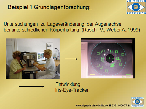
- Studies on the change of the axis of astigmatism from sitting to lying as a preliminary study for the development of an iris recognition system for the treatment of LASIK, (Rasch, Volker and Weber, Axel, 1999)
Implants
see also Cataract - Research
FAQ

What is refractive surgery?
In refractive surgery, refractive errors of the eye (the "refraction") are visually corrected by surgical interventions.
Are you a candidate for refractive surgery?
You only have to meet a few formal requirements for a refractive correction:
- Minimum age 18 years
- a stable refraction (eyeglass reading) for at least a year - this sometimes shifts the age limit by several years.
- Refraction values in the area of successful and secured correction options
- There should be no eye disease (eg cataract or glaucoma) or general illnesses (eg rheumatism). There is a special need for advice here.
- Diagnostics and treatment should not be done during pregnancy or lactation.
- For people with allergies, the operation should be placed in an allergy-free time.
But your personal, deliberate decision for this procedure is especially important.
A refractive correction is a surgical procedure and, like all interventions of this kind, it involves risks. The risks are low, however, as serious complications are extremely rare. However, they must be included in your considerations and our joint planning. The success of the correction depends primarily on the refractive features, the anatomy of your eye, but also on your expectations.
Which procedure for which ametropia?
After a thorough preliminary examination and detailed consultation, we will discuss with each patient the medically and individually most sensible treatment for him and his eyes.
Who bears the cost?
Refractive surgery is so-called desired medicine. The costs are generally not covered by the health insurance companies. However, there are special cases, especially for private insurance.
Possible complications:
As with any surgery, lens replacement can also cause complications during or as a result of surgery. We advise you on this, if such an action should be indicated to you.
What is behind "LASIK-Plus"?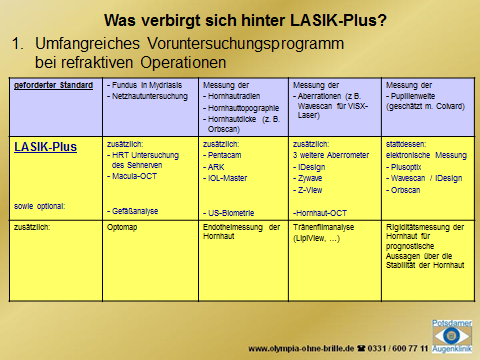


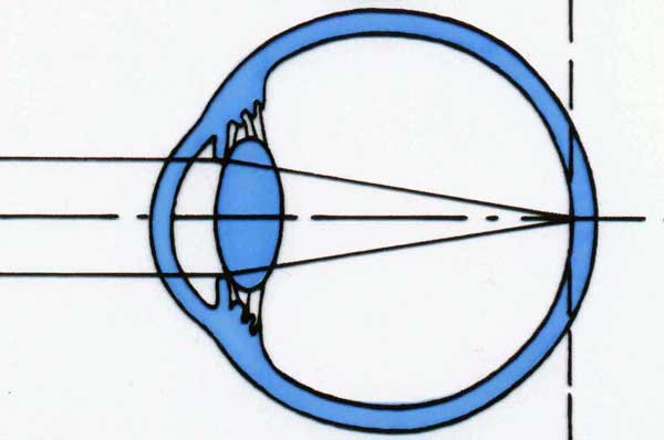 In the human eye there are three structures responsible for the refractive power of the eye:
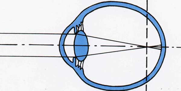 The short-sighted eye is relatively too long. Because of that, the light passing through the eye is "bundled" before hitting the retina. Far away objects don't appear sharp. The longer the eye, the higher the grade of myopia. People suffering from myopia can see objects that are close better than ones that are distant, which leads to the term "shortsightedness".
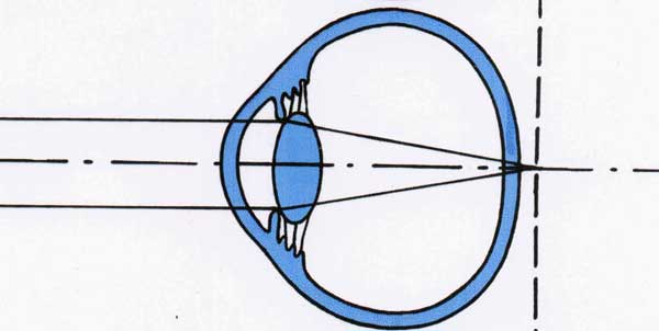 Hyperopic eyes are relatively too short. The light is theoretically bundled behind the retina, causing a blurred image. Younger people can balance the insufficient refractive power, because their lens will be temporarily curved only by looking into the distance. The focus will be set further to the front and onto the retina.
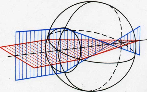 The bulge of the cornea in most human eyes is equally comparable to the sruface of a sphere. Whenever the cornea is warped stronger or less strong, objects will appear blurry and distorted. Because of this a perfectly round soccer ball can appear to look like a rugby ball and a dot may be seen as a bar.
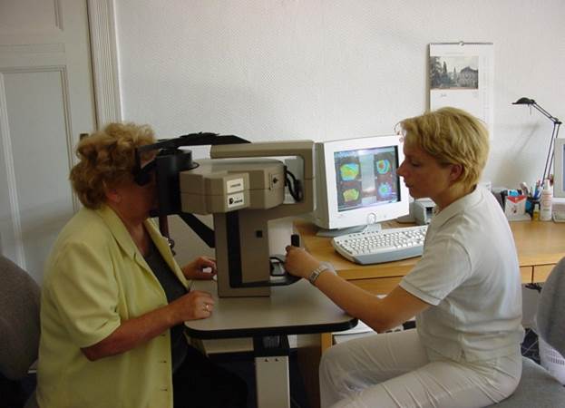 topography of cornea (e.g. Orbscan II / Pentacam)
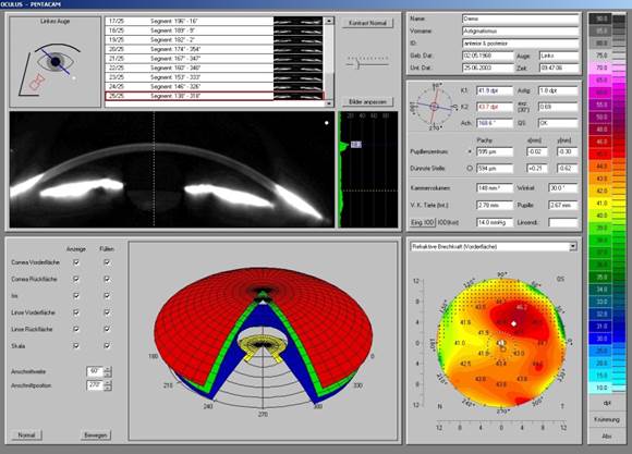 Pentacam
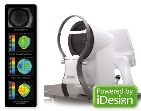 Aberrometry
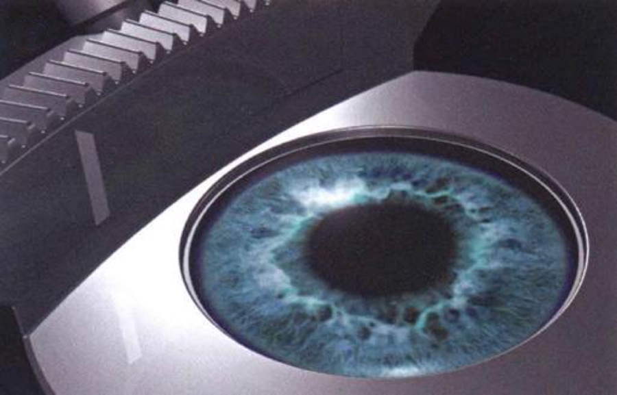
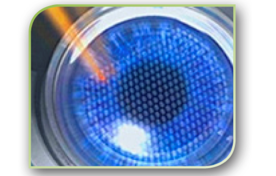
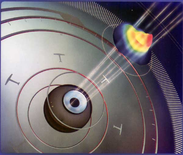 Other technical features of the VISX Star S4 ™ IR also help to improve surgical results. By means of iris registration (IR), the finest movements of the eye can be corrected under the excimer laser. Since all wavefront measurements are made while the patient is sitting, but treatment is done while the patient is lying down, cyclorotations (twisting of the eye) may occur (J. Stevens, ASCRS 2005). These movements can be compensated for by means of a complex iris registration, which is particularly important in treatments of astigmatism.
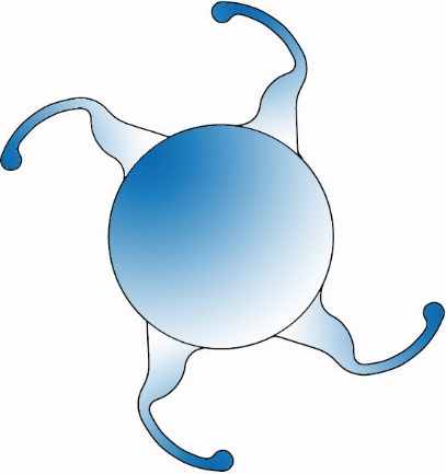 This involves replacing the natural lens with a new, artificial one with correction of ametropia. The clear body lens is removed similarly to the operation of cataract and replaced by an artificial one. The previously calculated lens should compensate for the refractive error.
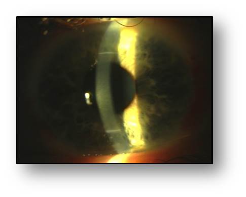 In all treatment options, a co-correction of existing astigmatism is possible. It is to be examined on a case-by-case basis which role the cornea and eye lens play.